
Neurosurgery refers to the clinical field that addresses surgical management of conditions affecting the brain, spinal cord, peripheral nerves, and associated structures. It encompasses procedures intended to diagnose or treat structural, vascular, degenerative, neoplastic, and traumatic conditions of the central nervous system. Practitioners in this field coordinate imaging interpretation, surgical planning, operative execution, and post-procedural follow-up to address anatomic or physiologic disruptions within neural tissues, using a range of open and less-invasive techniques depending on anatomy and clinical context.
Procedural approaches in this area typically involve multidisciplinary input, including neurosurgeons, neuroradiologists, anesthesiologists, and rehabilitation specialists. Preoperative evaluation often focuses on imaging, functional assessment, and risk stratification to match a surgical plan to a patient’s condition. Intraoperative methods and postoperative pathways vary by target (intracranial versus spinal), and the practice generally balances aims of symptom control, structural stabilization, and preservation of neurological function while acknowledging perioperative risks and recovery trajectories.
Selection among these and related procedures typically follows a diagnostic framework that may include clinical examination, cross-sectional imaging, and an assessment of functional goals. For example, an intracranial lesion that produces mass effect and progressive symptoms may be considered for resection when imaging and functional mapping indicate a reasonable risk–benefit balance. For spinal conditions, instability or neural compression often informs whether decompression alone or decompression with fusion is contemplated. These determinations frequently involve multidisciplinary case review and patient-centered discussion rather than a single metric.
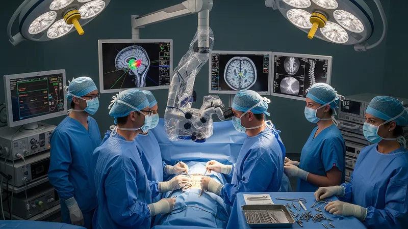
Imaging and intraoperative guidance tools often play a central role in modern neurosurgical practice. High-resolution MRI, CT, and angiographic studies may be used to define anatomy and plan surgical corridors. Intraoperative neuronavigation systems and real-time electrophysiological monitoring can provide additional spatial and functional context, which may influence approach selection and extent of intervention. These adjuncts are intended to support clinical judgment and may help reduce uncertainty during complex procedures.
Operative techniques range from traditional microsurgical exposure to minimally invasive tubular or endoscopic corridors, and from open spinal instrumentation to percutaneous fixation. Each technique carries procedural trade-offs that surgeons and teams typically weigh, such as tissue disruption, visualization, and anticipated recovery course. Endovascular methods may be chosen for vascular lesions when intraluminal access offers a lower anatomical disruption compared with open exposure. Procedural choice often reflects anatomy, underlying pathology, and available expertise.
Post-procedural frameworks commonly integrate early assessment of neurological status, pain management strategies, wound care, and rehabilitation planning. Discharge planning may consider mobility, home support, and the need for outpatient or inpatient rehabilitative services. Outcome measurement is usually multidisciplinary and may include serial imaging, functional scales, and longitudinal follow-up to monitor for complications and to guide rehabilitation intensity. These pathways are adapted to individual clinical contexts rather than standardized in every case.
In summary, the concept covered here addresses surgical interventions for brain and spine conditions through a sequence of evaluation, planning, operative technique, and follow-up. The examples provided illustrate representative procedural categories and the broader considerations that typically inform their use. The next sections examine practical components and considerations in more detail.
Different surgical approaches are applied depending on anatomic location and pathology. For intracranial lesions, open craniotomy remains a common route that may be tailored by skull flap placement and cortical mapping. Minimally invasive skull-base approaches and endoscopic corridors can be used for selected midline or ventral lesions to limit brain retraction. For the spine, posterior decompression, anterior column access, lateral approaches, and combined strategies provide options for addressing neural compression or instability. Approach selection often considers tissue exposure needs, the vascular relationship of lesions, and anticipated postoperative function.
Minimally invasive techniques have grown as an option in many centers and may affect hospitalization length and recovery milestones in some patients, although applicability varies by lesion type and surgeon experience. For example, tubular retractor systems can permit targeted lumbar decompression with smaller soft-tissue exposure, whereas open posterior approaches may be preferred for multilevel instability requiring complex instrumentation. Endovascular approaches are an alternative for vascular pathology when intraluminal access is anatomically feasible and imaging suggests an acceptable route.
Surgeons typically review imaging with multidisciplinary colleagues to assess approach feasibility and to anticipate intraoperative challenges, such as vascular encasement or proximity to eloquent cortex or nerve roots. Surgical planning may also incorporate functional mapping to preserve language, motor, or sensory pathways when operating near critical regions. Considerations about the patient’s overall health, prior surgeries, and goals of care inform whether a more conservative or more aggressive route is selected.
When comparing approaches, teams may weigh short-term recovery considerations against long-term structural aims, such as stability after spinal fusion or extent of resection for intracranial lesions. Each choice involves trade-offs in visualization, tissue handling, and potential need for staged procedures. Readers should note that procedure selection is individualized and that published comparative data often describe averages or trends rather than deterministic outcomes for any single patient.
Preoperative planning commonly begins with detailed imaging to characterize lesion size, location, and relation to surrounding structures. MRI provides soft-tissue contrast useful for tumor and cord pathology, while CT better depicts osseous detail relevant to spinal instrumentation or skull base anatomy. Vascular studies such as CTA, MRA, or catheter angiography may be necessary for vascular lesions. These data sets are often integrated into planning workstations and neuronavigation systems to visualize surgical corridors and to estimate resection margins or fixation trajectories.
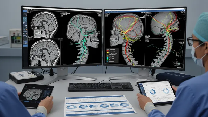
Functional imaging and mapping may be used in selected cases where preserving neurologic function is a priority. Functional MRI and tractography can illustrate likely pathways for language or motor tracts and may inform whether awake mapping or intraoperative stimulation is needed. Risk assessment also includes medical comorbidity evaluation, anticoagulation management, and considerations about prior radiation or surgery that may affect tissue planes and healing potential. Multidisciplinary planning meetings can help align goals and expectations.
Three-dimensional modeling and simulation are increasingly used for complex cranial and spinal deformity planning; these techniques may facilitate rehearsal of osteotomies or hardware placement in some centers. For endovascular planning, digital subtraction angiography provides fine vascular detail to guide catheter-based strategies. Planning frameworks typically prioritize patient safety and functional preservation, using imaging to reduce uncertainty about critical relationships and access routes before entering the operating room.
Informed consent discussions frequently reflect imaging findings and procedural alternatives, addressing potential benefits and limitations in nonabsolute terms. Documentation often includes anticipated hospital course and follow-up imaging plans to monitor for residual disease, hardware position, or vascular recurrence when applicable. Such structured planning aims to align the surgical strategy with measurable anatomic goals and the patient’s clinical priorities.
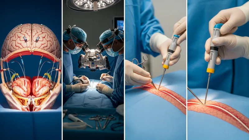
Operative technique selection is linked to the targeted pathology and the planned approach. Microsurgical techniques use high-magnification optics and fine instrumentation for intracranial and intradural spinal work, emphasizing precise tissue dissection and hemostasis. Endoscopic tools can allow visualization through smaller openings, while open approaches may be preferred when broad exposure or complex reconstruction is needed. Spinal instrumentation typically requires careful alignment and secure fixation with screws, rods, or cages designed to maintain stability and promote fusion in appropriate contexts.

Intraoperative neurophysiological monitoring (IONM) is commonly used to provide real-time data about neural function during procedures that risk motor or sensory pathways. Modalities such as somatosensory evoked potentials, motor evoked potentials, and electromyography may be applied selectively depending on the surgical target. Monitoring outputs are interpreted cautiously by clinicians, and alerts are integrated with surgical maneuvers to reduce the likelihood of iatrogenic injury. Monitoring is an adjunct that may inform intraoperative adjustments rather than guaranteeing outcomes.
Anesthesia and perioperative management strategies are tailored to the procedure and monitoring needs. Awake craniotomy protocols may be used when intraoperative functional testing is required, while general anesthesia with neuroprotective strategies may be chosen for extensive resections. Blood loss management, temperature control, and fluid balance are components of intraoperative care that typically receive coordinated attention from the surgical and anesthesia teams. These factors can affect immediate postoperative recovery and are considered during preoperative planning.
Surgical teams often maintain checklists and sterile-field procedures to minimize infection risk and to ensure equipment readiness, including navigation calibration and availability of vascular or graft materials. In complex cases, staged procedures may be considered to manage surgical risk and to allow for reassessment between stages. These intraoperative considerations are practical elements that often influence operative duration, resource needs, and postoperative pathways.
Immediate post-procedural care commonly emphasizes neurological monitoring, pain management, and prevention of complications such as infection or thromboembolism. Patients may be observed in a high-acuity setting for a variable period before transition to ward-level care. Early mobilization and respiratory care are often prioritized to reduce secondary complications. Wound inspection and basic functional screening may begin soon after surgery to identify evolving issues that warrant earlier intervention or imaging.

Rehabilitation planning is typically individualized and may include physical therapy, occupational therapy, and, when indicated, speech-language pathology. The intensity and duration of rehabilitation may vary based on the procedure and baseline function; some patients engage in short-term outpatient therapy, while others may require prolonged, multidisciplinary inpatient rehabilitation. The aim in these programs is to maximize functional recovery and adaptive strategies rather than to promise specific outcomes.
Follow-up imaging and clinical assessment schedules are often used to monitor for residual disease, hardware position, or vascular recurrence. Imaging timelines vary by pathology; for example, early postoperative imaging may verify resection extent or hardware placement, while later studies assess healing and longer-term stability. Clinical follow-up typically includes serial neurologic exams and review of functional progress, with adjustments to therapy plans made as recovery trajectories become clearer.
Long-term considerations may include management of chronic pain, activity modification, and secondary prevention of further neurologic events where applicable. Communication across surgical teams, primary care, and rehabilitation providers helps coordinate ongoing care and supports reintegration into daily activities. Readers should note that timelines and specific interventions are individualized, and multidisciplinary coordination often shapes the rehabilitation pathway.