
Artificial intelligence in breast cancer treatment refers to computational methods that analyze clinical data to assist clinicians in diagnosis, planning, and ongoing care. These methods typically use machine learning models trained on imaging, pathology slides, electronic health records, and genomic data to identify patterns that may be difficult for a human reader to detect consistently. The goal of these systems is informational and supportive: to provide probabilities, highlight areas for review, or summarize complex datasets so care teams can integrate that input with clinical judgment.
Applications of these computational approaches span screening workflows, diagnostic interpretation, treatment selection, and longitudinal monitoring. In imaging, algorithms may flag regions of interest on mammography or tomosynthesis images. In pathology, automated image analysis may quantify cellular features or suggest tissue classifications. Predictive models that combine multiple data types can provide risk estimates or likely trajectories, which clinicians may consider alongside established guidelines and patient-specific factors.
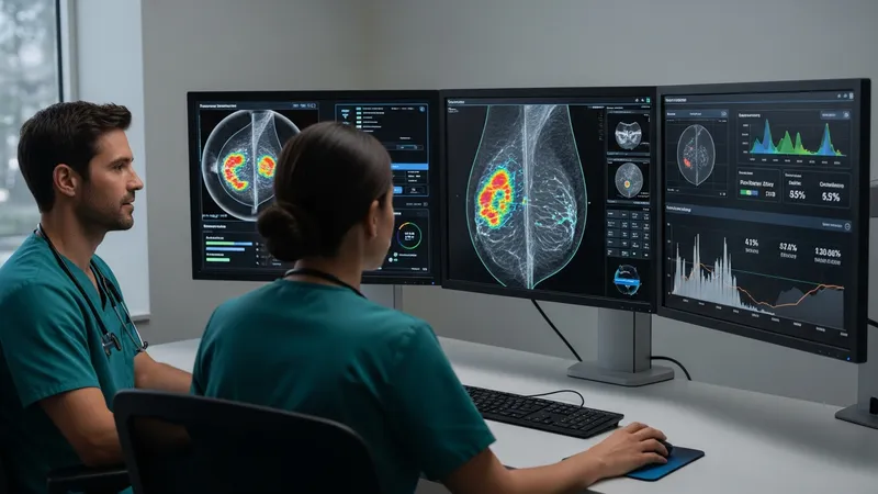
When comparing these example tool types, it is important to view them as complementary components rather than replacements for clinical expertise. Imaging detection software often focuses on sensitivity for certain lesion types but may vary by dataset and device. Digital pathology algorithms can standardize some quantitative measures but typically require human pathologist confirmation. EHR-integrated models that incorporate multiple data sources may improve situational awareness but are sensitive to the completeness and formatting of input data. Transparency about training data and intended use cases is a practical evaluation point.
Data quality and representativeness are central considerations for these computational methods. Models trained on limited or homogeneous datasets may perform differently when applied to diverse patient groups or imaging equipment. Validation studies often report performance on specific cohorts; readers should note that published results may reflect controlled datasets. Independent external validation and peer-reviewed evaluations are commonly cited practices to understand how a given method may behave in broader clinical settings.
Regulatory and implementation contexts influence how these systems are adopted in clinical practice. Depending on jurisdiction, some algorithms intended to inform diagnosis or treatment planning may require regulatory review or clearance. Operational integration with imaging systems, laboratory workflows, and electronic records typically involves technical and clinical governance steps. Institutions often pilot new tools, assess workflow impact, and define oversight mechanisms to ensure the output is used as intended and documented appropriately.
Ethical and practical considerations such as interpretability, data privacy, and clinician oversight are integral to responsible use. Interpretability features may help clinicians understand which inputs influenced an output, while privacy protections govern how patient data are stored and processed. Ongoing monitoring, periodic re-evaluation of model performance, and mechanisms for reporting unexpected behavior are often described in guidance documents on clinical AI deployment.
In summary, artificial intelligence approaches in breast cancer care encompass imaging analysis, pathology support, predictive analytics, and clinical decision support tools that may assist teams in diagnosis, planning, and monitoring. These methods typically augment information available to clinicians rather than dictate care, and their utility depends on data quality, validation, and appropriate oversight. The next sections examine practical components and considerations in more detail.
Imaging analysis and digital pathology are among the most visible AI applications in breast cancer workflows. Imaging algorithms often process mammography, tomosynthesis, ultrasound, or MRI to highlight areas that warrant focused review. Pathology platforms commonly analyze digitized slides to quantify features such as mitotic figures, cellular density, or biomarker staining intensity. These outputs can serve as structured inputs for multidisciplinary case review, where radiologic and pathologic findings are considered together. Care teams may view algorithmic outputs as one data layer alongside human interpretation and additional testing.
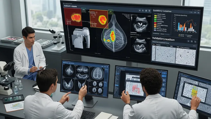
Performance differences across imaging modalities and lab processes can affect how an algorithm is used. For example, image acquisition settings, vendor equipment, and staining protocols can change input characteristics. Developers and institutions often report validation on specific scanner types or staining workflows, so implementers may run local verification studies. In research literature, reported improvements in sensitivity or specificity are typically context-dependent; therefore, local clinical validation and calibration are often described as prudent steps before routine reliance.
Interoperability between imaging systems, digital pathology viewers, and electronic records is an implementation consideration. Integration facilitates consolidated reporting and reduces manual transcription errors, but it may demand technical work such as mapping data fields and ensuring secure data transfer. Institutions sometimes prioritize staged deployments—starting with read-only or triage modes—so clinicians can compare algorithm output with standard practice before changing diagnostic pathways.
Operational governance around imaging and pathology AI generally includes labeling expected use cases, documenting performance metrics in real-world settings, and defining clinician responsibilities for review. Clinical workflows frequently specify that algorithm outputs require human confirmation and are to be interpreted in the clinical context. Reporting mechanisms may capture discordant cases to inform iterative model updates and maintain a record of decision rationale.
Risk assessment and predictive analytics combine demographic, imaging, pathology, and sometimes molecular data to estimate probabilities relevant to breast cancer screening and management. Models can be designed to estimate short-term likelihood of a malignancy on a current image, longer-term risk of developing cancer, or probabilities of recurrence after treatment. Such probabilistic outputs typically serve as part of a broader assessment, informing surveillance intensity, biopsy thresholds, or discussion of options rather than providing definitive conclusions.
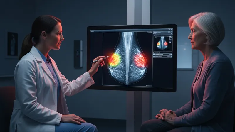
Model validity depends on the representativeness of training cohorts and the scope of input variables. Predictive tools trained on datasets reflecting certain age groups, ethnicities, or imaging practices may perform differently in other populations. Published work often emphasizes the need for external validation; some studies report modest improvements in risk stratification when multimodal data are combined. Users commonly interpret risk estimates as an additional piece of evidence and weigh them against clinical examination, family history, and patient preferences.
Transparency in model outputs can aid clinical interpretation. Outputs that include contributing factors or explainability markers may help clinicians understand drivers of a given risk score. This transparency can support discussions with patients about surveillance decisions and expected trade-offs. It also facilitates quality assurance efforts by revealing unexpected dependencies on a single input source or data artifact that might bias predictions.
Operational considerations for predictive analytics include updating models over time and monitoring for performance drift. As practice patterns, screening technologies, or population demographics change, models may require retraining or recalibration. Institutions interested in adopting such tools often set up monitoring plans that compare predicted versus observed outcomes and document any changes in workflow or decision thresholds that result from the model’s use.
AI approaches in treatment planning can contribute to therapy selection, radiation planning, and surgical guidance by summarizing data, suggesting potential targets for review, or automating time-consuming tasks. In radiation oncology, algorithms may assist with contouring volumes on imaging studies or estimating dose distributions. In multidisciplinary discussions, model-generated summaries that combine imaging, pathology, and prior treatment courses may help teams identify options to consider. Importantly, these systems typically provide informational outputs that clinicians review and adapt based on patient-specific factors.
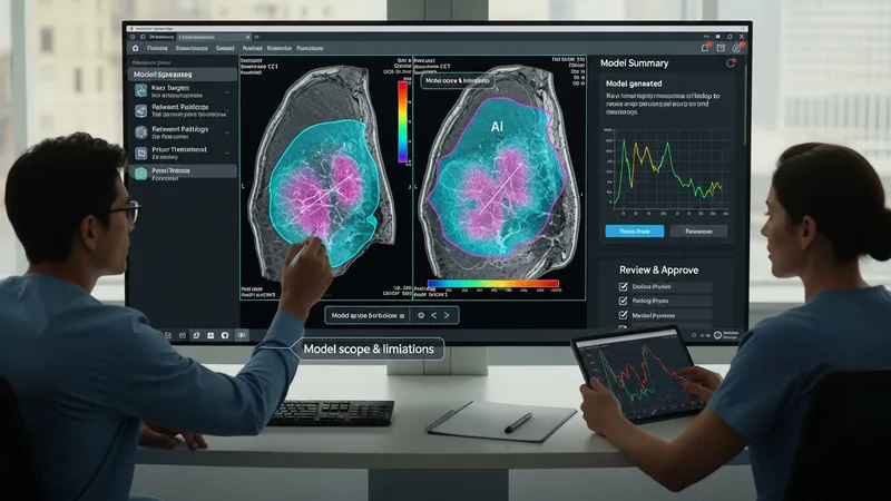
When applied to therapy planning, transparency about model scope and limitations is important. Algorithms trained to propose contours or dose plans are often validated on curated datasets and may require human modification for individual anatomy or comorbidities. Surgical planning tools that highlight likely tumor boundaries on images can aid preoperative assessment, but intraoperative decision-making continues to rely on direct visualization and surgeon judgment. Documentation practices usually specify that algorithm-assisted plans are reviewed and approved by qualified clinicians before implementation.
Cost and resource considerations can influence adoption of treatment-planning tools. Some approaches automate labor-intensive tasks and may free clinician time for other activities, while others require substantial technical integration and staff training. Institutions may conduct pilot evaluations to estimate implementation time, informatics needs, and potential workflow adjustments. Decisions to scale a tool often consider both technical benefits and resource implications for clinical teams.
Clinical evidence for therapy-support applications often includes retrospective comparisons and limited prospective studies; prospective controlled evaluations can be more challenging to perform but may provide stronger evidence about clinical impact. Stakeholders commonly look for peer-reviewed studies, technical documentation of training data, and local feasibility assessments when deciding how to incorporate such tools into routine planning workflows.
Beyond individual diagnostic or planning tasks, AI can support broader clinical workflows and longitudinal patient monitoring. Examples include automated triage queues that prioritize imaging or pathology cases for rapid review, natural language processing that extracts structured data from clinical notes, and remote monitoring systems that analyze patient-reported outcomes or device-derived signals. In research, these methods often accelerate cohort identification, feature extraction, and hypothesis generation for clinical studies.

Implementation in routine care usually involves multidisciplinary coordination among clinicians, informaticians, and compliance teams. Workflow changes often aim to preserve clinician oversight while reducing repetitive manual work. Monitoring of system performance in live settings is commonly established, with mechanisms to flag discrepancies or unintended effects. These governance measures help ensure that algorithmic assistance aligns with clinical goals and institutional policies.
From a research perspective, curated datasets and open-source benchmarks have facilitated comparative evaluations of algorithms. Researchers often report that reproducibility and dataset diversity are critical for assessing generalizability. Collaborative initiatives may promote shared standards for performance reporting and interoperability, which can support more informed adoption decisions by clinical teams and contribute to evidence accumulation about real-world impacts.
Practical considerations for monitoring and maintenance include updating models as new data accumulate, documenting versions used in clinical decision-making, and training staff to interpret outputs. Institutions commonly establish review cycles to assess whether algorithm use alters diagnostic or treatment patterns in expected ways and to determine whether additional validation or refinement is needed to maintain safe and informative integration into patient care.