
Handheld point-of-care ultrasound refers to compact, portable imaging devices that allow real-time sonographic assessment at the bedside or in ambulatory settings. These systems typically combine a small transducer with onboard or paired display software, enabling clinicians to visualize anatomical structures and physiological motion without the footprint of a cart-based unit. Devices in this category may use wireless connectivity to tablets or smartphones, and often emphasize rapid setup, battery operation, and simplified imaging presets intended for focused assessments rather than full diagnostic studies.
Key characteristics of these handheld systems include probe form factor, image processing algorithms, battery life, and the software interface for image display and storage. Some models use conventional piezoelectric transducers while others adopt silicon-based or single-probe architectures that attempt to cover multiple frequency bands. Data security, image export formats, and integration with local workflows can vary across manufacturers, and regulatory clearance for specific clinical uses is typically device- and jurisdiction-dependent.
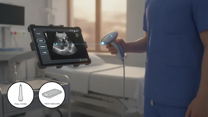
When comparing handheld units, image resolution, probe coverage, and software features often guide selection. Image quality can be sufficient for focused assessments such as cardiac window visualization, lung sliding, or limited abdominal surveys, while broader diagnostic studies may still rely on full-size systems. Connectivity options may include Wi‑Fi, Bluetooth, or wired connections to mobile devices. Storage and export support for DICOM or proprietary formats affects how images enter patient records. Regulatory approvals and intended use statements often define which clinical tasks a device may be used for in a given jurisdiction.
Workflow integration typically examines how a handheld ultrasound fits into triage, bedside assessment, and follow-up workflows. Some settings use these devices to augment physical exam findings quickly, potentially reducing time to initial assessment. Integration points include image annotation, automatic measurement tools, and cloud or local archiving. Institutions often consider data governance, user access levels, and compatibility with electronic health record systems when adopting handheld ultrasound into clinical pathways.
Device operation practices include probe handling, contact technique, and selection of imaging presets for different tissue types. Users may employ high-frequency linear presets for superficial structures like vessels and soft tissues, and lower-frequency phased-array or curvilinear presets for deeper abdominal or cardiac views. Battery management, probe disinfection protocols, and routine software updates are practical factors that may influence availability and image consistency across shifts. Training programs typically emphasize hands-on scanning and image interpretation competencies rather than formal device endorsement.
Clinical contexts where handheld ultrasound is often applied include focused cardiac exams, lung assessments for pleural motion, vascular access guidance, and limited abdominal checks for free fluid. These applications rely on focused protocols and may use simplified scoring or measurement approaches. Limitations include smaller display size, potential trade-offs in penetration or resolution, and the need to recognize when a comprehensive diagnostic study on a full-featured system is appropriate. Users should treat handheld imaging as an adjunctive assessment tool within a broader diagnostic workflow.
In summary, compact wireless ultrasound devices are designed to enable targeted, point-of-care sonographic assessment through portable transducers and mobile display solutions. They may streamline focused evaluations and offer connectivity features that support image sharing and archiving, while also presenting considerations for image quality, workflow integration, and governance. The next sections examine practical components and considerations in more detail.
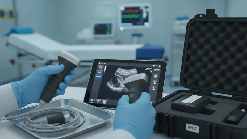
Physical design and portability determine where and how handheld ultrasound devices can be used. Many models aim for a balance between a lightweight transducer and sufficient battery capacity to support multiple scans per shift. Wireless probes that pair with mobile displays can reduce cable clutter and increase mobility, while tethered probe-and-tablet configurations may offer more predictable connectivity. Case size, weight, and available probe footprints often influence use in emergency departments, outpatient clinics, or field environments. Device durability and ingress protection ratings can also be relevant for use outside controlled imaging suites.
Ergonomics and user interface design can affect scanning efficiency and user fatigue during repeated exams. Probe handle shape, button placement, and the responsiveness of touch-screen or physical control options often shape operator preference. Mobile applications commonly provide exam presets, measurement tools, and image optimization controls; the intuitiveness of these interfaces may reduce time to diagnostic-quality images for routine focused exams. Heat dissipation, slip resistance, and ease of cleaning are additional design factors that may affect daily practical use.
Battery life and charging workflows are operational considerations that often influence deployment choices. Typical handheld units may support several hours of active scanning or a set number of examinations per charge; institutions sometimes maintain charging docks or rotating loaner devices to ensure availability. Battery performance may vary with screen brightness, wireless transfer activity, and the use of power-intensive software features such as continuous Doppler or high-frame-rate modes. Planning for charging cycles and spare devices can reduce downtime in high-volume settings.
Material and ingress protection ratings provide insight into a device’s suitability for varied environments. Some devices include water- and dust-resistance ratings intended to support routine cleaning and disinfection processes. Agencies or facilities may establish cleaning protocols aligned to device recommendations to balance infection control and device longevity. When selecting or deploying handheld ultrasound, matching device durability and maintenance patterns to the intended clinical environment can support sustained operation and reduce unexpected interruptions.
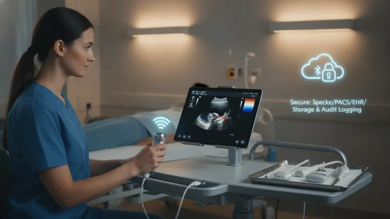
Connectivity options shape how handheld ultrasound images are displayed, stored, and shared. Devices that pair wirelessly with mobile phones or tablets often rely on Wi‑Fi or Bluetooth protocols; some also support direct wired connections for faster image transfer or charging. Cloud-based storage and vendor-hosted archives may be available, but local on-device storage and export to institutional PACS or EHR systems are common integration considerations. Data encryption, user authentication, and audit logging are technical factors that institutions often evaluate for secure image handling.
Imaging capabilities vary by probe technology and onboard processing. Some handheld probes combine multiple frequency bands into a single transducer, enabling a range of superficial to deeper tissue views, while others use dedicated probes for linear or phased-array imaging. Software features such as harmonic imaging, speckle reduction, and basic measurement tools may improve visual interpretation for focused exams. Color flow or pulse-wave Doppler capabilities are less common in the smallest devices but may be present in higher-tier handheld systems.
Image quality and frame rate often affect the range of clinical tasks that are appropriate for a handheld unit. For dynamic assessments like cardiac motion or lung sliding, temporal resolution can be important. For vascular access or soft tissue work, spatial resolution at superficial depths may be prioritized. Operators typically consider the balance between penetration depth and resolution when selecting presets; device documentation frequently outlines recommended exam types and expected image performance characteristics.
Interoperability with existing clinical IT systems is a practical consideration for deployment. Support for standard formats such as DICOM, HL7 interfaces, or secure file transfer protocols can influence how efficiently images and reports are incorporated into patient records. Device vendors may provide APIs or integration toolkits to assist with automated workflows, but institutions often require validation of data flow, retention policies, and compliance with applicable privacy regulations prior to routine clinical use.
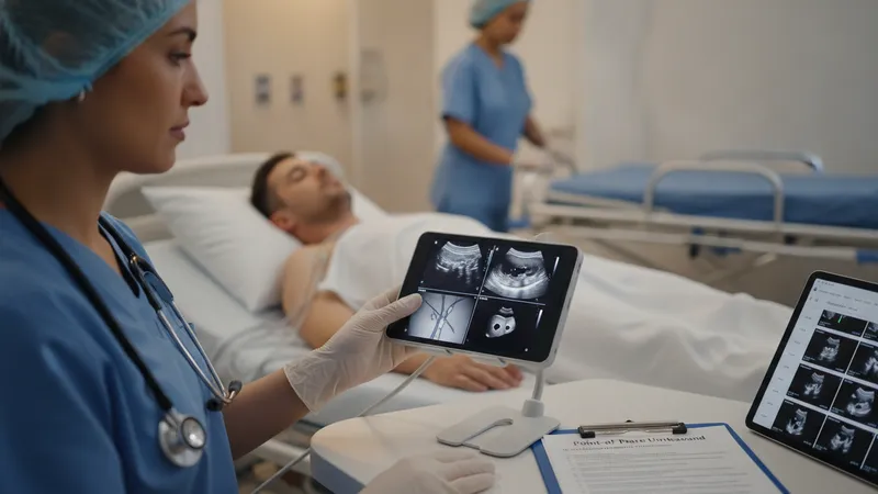
Handheld ultrasound devices are frequently used for focused, point-of-care applications where rapid visual information augments a physical assessment. Common use cases include limited cardiac windows for gross ventricular function assessment, lung evaluations for pleural motion or effusion, guidance for vascular access, and quick abdominal checks for free fluid. In many settings, handheld devices act as an extension of the physical exam and may shorten time to initial assessment; however, they typically complement rather than replace comprehensive diagnostic imaging when detailed evaluation is required.
Workflow integration addresses when and how these devices are used during patient encounters. Protocols may define specific indications for handheld scans, documentation standards for image capture, and escalation pathways when findings suggest additional imaging. In outpatient clinics, handheld scans can support same-visit decision discussions by providing immediate visual context. In emergency or transport settings, rapid assessments that identify gross abnormalities may influence triage and immediate management planning, though confirmatory imaging on full systems may be needed for definitive evaluation.
Training and competency frameworks influence safe and effective use in clinical practice. Educational programs for handheld ultrasound often focus on probe handling, basic image acquisition, and interpretation of common focused views. Competency assessment may include supervised scanning, review of stored images, and alignment with institutional scanning protocols. Ongoing quality assurance activities—such as periodic image audits or peer review—may help maintain consistent image quality and appropriate use across teams or departments.
Operational policies can include device assignment, sterilization or disinfection methods, and data governance measures. Facilities commonly document acceptable use cases, image retention timelines, and responsibilities for software updates or maintenance. Clear documentation and training expectations help ensure that handheld imaging supports clinical workflows in a way that is consistent, auditable, and aligned to broader imaging governance frameworks without implying diagnostic replacement of conventional imaging studies.
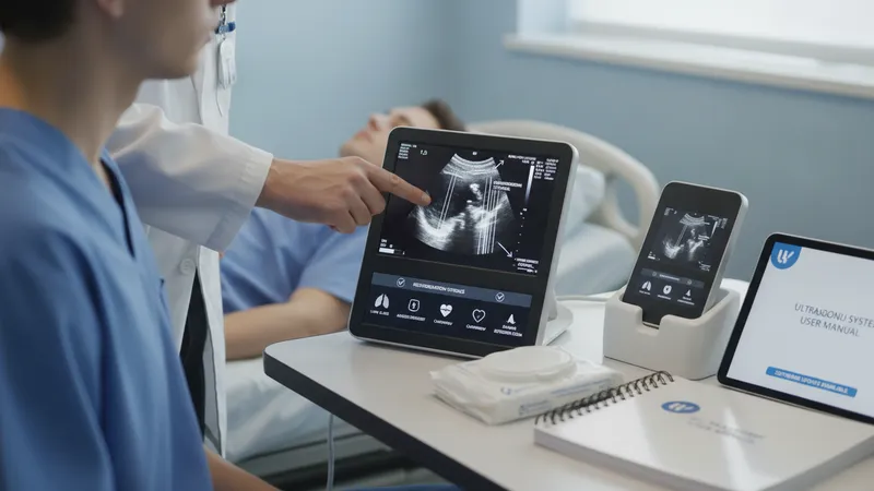
Effective operation of handheld ultrasound relies on structured training that emphasizes focused imaging protocols and recognition of common artifacts. Short, procedure-oriented training modules may cover insonation angles, probe pressure, and typical artifact patterns such as reverberation or shadowing. Supervised practice sessions and image review with experienced users often help new operators translate textbook views into reliable bedside images. Training typically frames handheld imaging as an adjunct that provides rapid visual information rather than a definitive diagnostic report.
Maintenance and device lifecycle considerations include software update policies, warranty coverage, and routine calibration or performance verification. Regular software updates may add functionality or improve image processing; facilities often schedule updates to minimize disruption. Cleaning and disinfection protocols should align with manufacturer guidance and institutional infection control standards to ensure patient safety while preserving device integrity. Planning for spare devices or service agreements can reduce downtime in high-utilization environments.
Quality assurance measures help ensure consistent image quality and appropriate use. Common practices include image logging, peer review sessions, and periodic audits comparing handheld findings with subsequent full studies when available. These activities may identify gaps in technique, interpretation, or workflow that can be addressed through targeted training or protocol adjustments. Documentation standards for saved images and brief structured reports assist in maintaining a traceable record of bedside assessments.
Adoption planning often assesses clinical needs, training resources, and data governance before routine deployment. Considerations may include identifying clinical champions for education, mapping connectivity pathways to the institution’s record systems, and defining permissible use cases aligned with regulatory guidance. By treating handheld ultrasound as a focused assessment tool with defined boundaries and oversight, organizations can integrate its capabilities into patient care pathways while maintaining clear expectations for when comprehensive imaging is required.